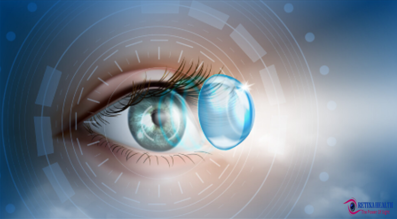
The human eye lens is a clear, curved structure that focuses light onto the retina at the back of the eye. It is similar to the lens of a camera.The lens of the eye is a relatively transparent structure, made of proteins and fluids.Crystalline proteins give the eye lens its clarity and transparency.
It is the part that focuses and transmits light behind the iris , where sensors in the cornea convert it into the retina of the visual data. The shape of the lens is a curved structure that’s embedded deep within your eye (or camera). It absorbs light and bends it to converge at a single point behind it. This focuses the light for the sensors at the back ,whether that’s camera film, digital sensors or, in your eye, the retina.
Lens Function:-
The eye lens works much like a camera lens absorbs , bending and focusing light to produce a clear image. Light rays enter the eye through the pupil, focusing them onto the retina. The crystalline lens is a convex lens that creates an inverted image focused on the retina. The lens changes its shape automatically to focus on objects that are close or far away. It can make itself flatter or rounder to bend incoming light from different distances toward a single point. The brain flips the image back to normal to create what you see around you. In a process called accommodation, the elasticity of the crystalline lens allows you to focus on images at far distances and near with minimal disruption.The lens provides about 30% of your eye’s focus; your cornea provides the other 70%. As you age, the lens may become weaker or damaged. Since the lens changes shape to focus on images near or far, it can grow weaker and may not work as well later in life
Lens Structure:-
The human eye lens is located behind the pupil , which is the dark spot in the middle of your iris. Iris is the coloured part of your eye ,suspended from the eye wall by small zonular fibers called zonules. The pupil is an opening that lets light into your eye. The iris controls the size of the opening and the amount of light coming in. Light passes through your pupil to the eye lens, which focuses it onto the retina behind it. This makes your eye lens the second-to-last layer in your eye. The lens is a clear, curved disk that sits behind the iris and in front of the vitreous of the eye. It is the part of the eye that focuses light and images from the outer world, bending them onto the retina. The human eye lens actually has no direct blood or nerve connections. Instead, it relies on aqueous humor ,the clear fluid between the lens and the cornea, to provide it with energy and carry away waste products.
The lens of your eye is made up of structural proteins called crystallins. This is why it’s sometimes called the “crystalline lens .The crystalline lens is a clear, biconvex layer of the eye that is made of collagen and protein.As much as 60% of the lens mass is made up of proteins, it has the highest concentration of proteins of almost any tissue in your body. proteins give the human eye lens its transparency and focusing power .Mature crystallins have no nucleus or organelles .Lens lose them as they mature But having no nucleus or organelles also prevents the cells from reproducing . This adds to their clarity and transparency.Four structures make up the crystalline lens:
- Capsule
- Epithelium
- Cortex
- Nucleus
Throughout your life, new cells continue to grow at the outer edges of the circle, while the older cells compress toward the center. Eventually, the older cells at the center begin to show wear and tear.The lens grows as you age, weighing about 65 milligrams at birth, 160 milligrams by age 10, and 250 milligrams by age 90.
Lens Development :-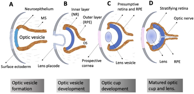
The human eye lens growth is a continuous process that occurs throughout a person’s life, forming sutures and a gradient refractive index.This growth occurs through the addition of new lens fibers to the outer layer (cortex) of the lens. Lens growth leads to changes in lens size, shape, and focusing ability. It is colourless at birth, but gradually becomes more yellowish with age. The lens grows as you age, weighing about 65 milligrams at birth, 160 milligrams by age 10, and 250 milligrams by age 90 .Age-related changes in lens growth contribute to presbyopia. There are two Phases of Growth:
- Rapid prenatal development: The lens grows rapidly during fetal development that ends shortly after birth.
- Slower postnatal development: After birth lens growth slows down but continues throughout life. A linear growth phase that adds 1.38 mg/year to the lens’s wet weight.
Conditions And Symptoms Of Lens:-
These two are common conditions in human eye lens.
- Cataracts
- Presbyopia
Cataracts:-
Cataracts are cloudy areas that form on the lens of your eye. Your lens is a clear, flexible structure made mostly of proteins (crystallins). As you get older, the proteins in your lens break down, forming cloudy patches that affect your vision.You may feel as if you’re looking at the world through a dirty window. Over time, your vision gets worse. You may have a hard time carrying out routine tasks. Age-related cataracts is the most common form of the condition But you don’t have to live with fading vision. Ophthalmologists can do surgery to remove the cataracts and restore your vision.
(Babies can be born with cataracts related to genetic disorders, and this is one way to recognize them.)Aging and environmental factors like sunlight eventually take their toll on your eye lens, particularly the older crystallin cells in the center. When these cells start to break down, they lose some of their transparency and become cloudy. This is what age-related cataracts.
Types of cataracts:-
There are many types of cataracts some are explain here.
- Pediatric cataracts: Pediatric cataracts affect babies and children. Babies may be born with cataracts (congenital), or the cataracts may form sometime after birth. Pediatric cataracts typically run in families, but they can also happen due to eye injuries or other eye conditions. Babies and children with pediatric cataracts need prompt treatment to prevent problems like amblyopia (lazy eye).
- Traumatic cataracts: These cataracts form when something injures your eye. Treatment for this type is more complicated because structures around the lens may also need repair.
- Secondary cataracts: These are cloudy patches that form on your lens capsule, or the membrane that covers your lens. Another term for this condition is posterior capsular pacification. It’s a common but easily treatable complication of cataract surgery.
Presbyopia:-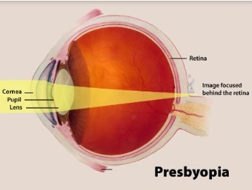
Presbyopia is the medical term for your eye becoming less flexible and losing its focusing ability. This causes me to have trouble focusing on things close-up. Presbyopia is essentially farsightedness (hyperopia) occurs as your eye’s natural lens grows less flexible with aging.Presbyopia generally starts to develop around age 40 and gets worse until your mid-60s . When your lens starts to become less flexible and loses its focusing ability, you’ll start to have trouble focusing on things close-up. Presbyopia is essentially farsightedness (hyperopia) that happens with aging. You might find yourself holding reading materials farther away from you to read. Or you might just notice that your eyes get tired from reading or doing close work more easily than they used to. You’ll notice yourself holding reading materials farther away from you to read other close-up tasks that are harder than they used to be. You might need to hold your book or phone out at arm’s length to see the words clearly. yourself holding reading materials farther away from you to read . You may also have symptoms like headaches or sore, tired eyes. z Presbyopia is part of the natural aging process, and it’s not a disease. It’s a common type of refractive error that eye care specialists can easily correct with glasses, contacts or surgery.Presbyopia is very common. Globally, about 1.8 billion people had presbyopia in 2015. Researchers estimate this number will rise to 2.1 billion by 2030.
Treatment of presbyopia:-
Depending on your health, lifestyle and preferences,following methods are used to correct presbyopia:
- Eyeglasses.
- Contact lenses.
- Surgeries.
- Eye drops.
Eyeglasses:-
Whether or not you’ve been wearing eyeglasses for other vision issues, now may be time to switch to a more comfortable type for your changing eyes.
- Reading glasses (readers): These are ideal for people who don’t have nearsightedness, farsightedness or astigmatism. You can buy readers from the store without a prescription (but it’s a good idea to ask your provider for the magnification power you need). You can also choose to get prescription reading glasses tailored to your eyes.
- Bifocals: Often prescribed for presbyopia, bifocals are glasses that have two different prescriptions in one spectacle lens. The upper part of the lens has a distance prescription, while the smaller, lower part has a prescription to help you see objects up close.
- Trifocals: Trifocals have three lenses: one each for seeing close-up, in-between and far away.
- Progressives: Progressives are multifocal lenses, similar to bifocals, but with a more gradual shift between the prescriptions. Many people choose progressives when they don’t want a visible line on their glasses.
- Office progressives: These glasses are designed for doing near work in the office, like computer work or writing. When you get up from your desk, you remove them so you can see into the distance.
Contact lenses:-
There are a variety of contact lenses that can help you see better with presbyopia.
- Bifocal contact lenses: A true bifocal lens helps you with just two focal points, usually near and far. They come in soft or hard (gas-permeable) materials.
- Multifocal contact lenses: Multifocal lenses are similar to bifocal lenses, and people often use the terms interchangeably. But a multifocal lens can include more than two focal points, including the in-between zone of about three feet. They also come in soft or gas-permeable versions.
- Monovision contact lenses: With a set of monovision contacts, one eye wears a lens that helps you see objects at a distance, while the other eye wears a lens that helps with near vision. Your brain gradually adapts to this method, but it can take up to two weeks for it to feel comfortable to you.
- Modified monovision contact lenses: With modified monovision, you wear one lens for either near or far vision. In the other eye, you wear a multifocal lens that helps you see at all distances.
Surgeries:-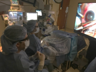
The following three laser surgery procedures correct presbyopia by using monovision (one eye corrected for distance, the other corrected for near vision):
- LASIK surgery: Laser in-situ keratomileusis (LASIK) is a popular surgical approach to correct vision in people who are nearsighted, farsighted or have astigmatism.
- PRK surgery. You may be a good candidate for a photorefractive keratectomy (PRK) procedure if you have moderate to high nearsightedness, farsightedness and/or astigmatism.
- SMILE surgery: With a small-incision lenticule extraction (SMILE) procedure, your surgeon uses a very precise laser to create a disc-shaped piece of tissue inside your cornea that they remove through a small incision.
Eye drops for presbyopia:-
Eye drops are a good option for some people with presbyopia. (Pilocarpine eye drops) make your pupil smaller to improve your depth of focus and give you clearer close-up vision. These are the first eye drops the U.S. Food and Drug Administration (FDA) has approved for presbyopia. Red eyes and headaches are the most common side effects, and you may also have trouble with night vision.

