The Iris Eye Color Palette is the colored tissue around the pupil, a ring-shaped membrane in your eye, located behind the cornea and in front of the eye. The iris is the eye part that makes up your eye color. It plays a critical role in overall health, controlling the amount of light that enters the eye and reaches back to the retina. The iris is a 12-mm diameter structure.
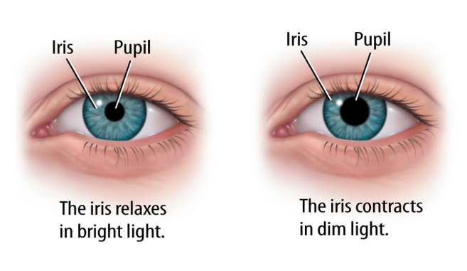
The iris plays a vital role in regulating the amount of light that can enter the retina. A circular muscle with a hole in the middle, the iris contracts and expands to control the amount of light that gets into the pupil. The iris has two tiny muscles that cause the pupil to contract and dilate. When outdoors, the pupil contracts and the iris expands to protect too much light from entering the eye. In low-light situations, the pupil opens wide, and the iris becomes smaller to let in as much light as possible, making it easier to see in low light.
The Iris Structure:-
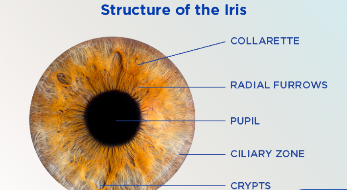
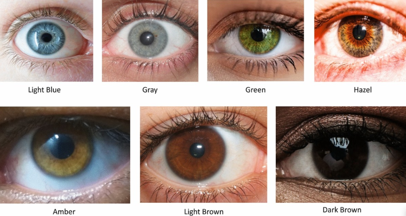
The Iris is present behind the cornea and above the eye lens. It originates from the ciliary body. The thick anterior portion of the choroid layer of the eye, which is known as the ciliary body extends further forming the iris It is a pigmented structure and the color depends on the amount and nature of the pigment present in it The typical eye color can be hazel, brown, grey, blue and green. The quantity of melanin pigment determines the color of the iris. The iris structure comprises three components: color, texture, and muscles.
Color: The iris gets its color from melanin, the pigment that determines skin and hair color. The amount of melanin determines eye color:
- High melanin: Brown eyes
- Moderate melanin: Hazel or green eyes
- Low melanin: Blue eyes
- Little to no melanin: Very rare cases of pink or red eyes (often associated with albinism)
Texture: The iris has a unique texture with patterns of ridges, furrows, and crypts. These patterns are so distinct that they can be used for biometric identification (iris scanning).
Muscles: The iris contains two main muscles:
- Sphincter pupillae: A circular muscle that constricts (narrows) the pupil in bright light.
- Dilator pupillae: Radial muscles that dilate (widen) the pupil in dim light.
The Iris Layers:-
The iris is the part of the eye that is a colored tissue. It is a circular muscle with a hole in the middle, The iris contracts and expands to control the amount of light that gets into the pupil. The iris sits in the uveal tract, which is the eye’s middle layer. It is made up of the following parts.
- Stroma: The front pigmented fibrovascular layer. Stroma is made up of connective tissue and blood vessels.
- Pigmented epithelial cells: Iris pigment epithelium cell is Present at the back. It contains melanin granules and chromatophores that make up the eye color.
- Dilator and sphincter: These muscles expand and contract to control the amount of light that gets in.
The iris eye color palette sits in front of the lens within the coronal plane towards the front of the eye. Unbound in its middle to allow the pupil to change size, this structure is connected to the ciliary body—the part of the eye that produces the eye’s fluid (aqueous humor) and regulates contraction and constriction of the iris. The iris splits the space between the cornea and lens into anterior and posterior chambers. The anterior chamber is bound by the cornea, while the posterior chamber connects with the ciliary bodies, zonules (a small anatomic band that holds the lens in place), and lens. Both chambers are filled with aqueous humor.
The Iris Function:-
The primary function of the iris is to regulate the amount of light that reaches the retina, the light-sensitive tissue at the back of the eye. The iris surrounds a small aperture called the pupil. The iris can narrow the pupil to prevent blurring caused by light rays moving in different directions. It does this by changing the size of the pupil, the black circle in the center of the iris. This pupillary light reflex is an involuntary response controlled by the autonomic nervous system. The iris plays a key role in regulating the amount of light that accesses the retina in the back of the eye. It contains two sets of muscles.
- Bright light: In bright light, the iris uses the circular muscle to decrease the size of the pupil and reduce the amount of light entering the eye.
- Dim light: In low light, the iris dilates to maximize the available visual information by contracting the radial muscles, widening the pupil to allow more light to enter.
Dilation and constriction of the pupil can also be influenced by physiological states, such as arousal and excitement. This structure performs the “accommodation reflex,” which is the eye’s involuntary ability to switch focus from objects that are nearby versus far away. This activity is regulated by the parasympathetic nervous system and involves:
- Changing the aperture (opening) of the pupil
- Changing the shape of the lens
- Convergence: the ability of the eyes to work together when looking at nearby objects.
Eye Color:-
Your eye color is determined by a combination of different pigments and saturation levels. Three main pigments are found in the iris:
- Melanin: A yellow-brown pigment that also determines skin tone, over half of the people in the world have brown eyes
- Pheomelanin: A red-orange pigment that causes red hair and is common in green or hazel eyes
- Eumelanin: A black-brown pigment that determines how intense or dark the iris is
Brown eyes have a higher amount of melanin, while blue eyes have very little pigment. The amount of pigment is often related to a person’s genes, skin type, and hair color.
Anatomical Variations:-
Anatomical variations are differences in the normal anatomy of the human body that occur between individuals, often without causing any health issues. These variations can include differences in the size, shape, or number of organs and structures. Anatomical variation plays an important role during diagnosis and treatment. Just like other body organs iris can have many anatomical variations, including:
- Aniridia in which the iris is incomplete or absent
- Low visual acuity (sharpness of vision)
- Photophobia (sensitivity to light)
- Degeneration of the macular and optic nerves (associated with processing visual information)
- Glaucoma can damage your optic nerve
- Color variation determines color according to the amount of melanin pigmentation
- A limbal ring a dark ring around the iris
- Genetic basis the variance in human eye color
- Dilator thickness can be thin or thick
- Cataracts (cloudy areas in the lens that affect vision)
- Changes in the shape of the cornea
- Involuntary eye movements (nystagmus)
This condition is associated with WAGR syndrome and Gillespie syndrome, two conditions characterized by disrupted organ function and intellectual disability.
Interesting Facts about iris:-
- Just like your fingerprints, iris patterns are unique to each individual, it work as biometric identity. Everybody has their own design, even identical twins have different patterns.
- The color of your iris depends on multiple genes and special pigment melanin in the body.
- The iris continuously changes the size of your pupil, In bright light, it shrinks your pupil to let in less light. In dim light, it makes your pupil bigger to let in more light.
- It also reacts according to your feelings like in astonishing condition or excitement, your pupils tend to become bigger.


