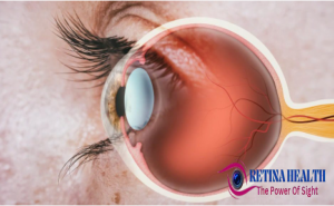
The Retina Human Light Sensor is the window of the World. Its intricate design and perceived functioning highlight the marvels of biology and evolution. Lets explore the wonder of the eye ,the eye inner canvas is one of the most valued and sensitive senses of organs. The pair of eyes present on the head allows us to see. It is the most important sense in our body while other senses like taste, hearing, smell, and touch. On closing the eye, we identify the objects to some extent by their smell, taste, and sound they make or by touch but identifying the color while closing the eye is impossible.  We perceive up to 80% of all impressions using our sight if other senses such as taste or smell stop working. It is the eyes that best protect us from danger Eye is almost spherical. It has an average diameter of 2.3 cm. A normal eye can see nearly 10 million different colors. An eye can see objects clearly that are between 25 cm and infinity. .20/20 vision is considered to be best but many people, mostly children, have better vision than 20/20. Human eye the eye is made up of several layers each playing a vital role in vision.
- Cornea
- Iris
- Pupil
- Lens
- Retina
- Optic nerve
- Vitreous humor
Unveiling the Retina Symphony Of Light And Sight:-
INTRODUCTION:-
The retina is like a film in the camera, a light-sensitive layer of the cell in the back of the eye; without it we can not see. It plays an important role in capturing light and converting it into electrical signals sent to the brain, via the optic nerve to process vision. You are Filled with millions of photoreceptor cells called rods and cones, the primary responsibility of the retina is to control how you see images. Rods allow you to see in dim lighting while cones help you see in daylight and decipher colors. Just like any part of the body, the retina can face many problems as well. All these problems can affect your vision health and in some special cases, lead to blindness. So make sure, taking care of your eyes is essential for healthy eyesight.
The word “Retina” comes from the Latin language “rete” which means “net”. The retina is the thin-sensitive layer of tissue at the back of the eye. It is the key connection between light and sight. This is the part of the central nervous system (CNS) and is present in brain tissue. Homo sapiens have two eyes and two retinas.
Key Components of the Retina:-
The main key components of the retina are mentioned here
Photoreceptor Cells:
Rods and cones, responsible for vision in low light and bright light.
Macula:
A small, specialized area in the center of the retina responsible for sharp, detailed central vision. It has a high concentration of cones.
Optic Nerve:
The bundle of nerve fibers that carries signals from the retina to the brain.
Conditions and Disorders:-
Several conditions can affect the retina some are:
- Age-related macular degeneration (AMD):
A common condition that damages the macula, leading to blurred central vision.
- Diabetic retinopathy:
Damage to the blood vessels in the retina caused by diabetes.
- Retinal detachment:
When the retina separates from the back of the eye.
- Retinitis pigmentosa:
A group of genetic disorders that cause progressive damage to the retina
Healthy Retina:-
In a healthy eye, light energy sent through the cornea of the eye and focused on the retina. The retina converts the energy into electrical signals that are transmitted through the optic nerve to the brain. Those electrical signals are then translated into images for you to understand. The macula is located in the center of the retina. A healthy macula will allow you to have clear central vision.
Damaged Retina:-
 Any damage to the retina or macula will affect how light energy is received and transferred to the brain. Central vision problems as well as overall blurriness, spots, and even blindness can occur when there is any deterioration of cells, retinal detachment, tears, puckers, or any type of damage to this sensitive area.
RETINAL LAYERS AND THEIR FUNCTIONS:-
The retina is n made up of three layers of nerve cells:
- Ganglion cells
- Bipolar cells
- Photoreceptor cells
Ganglion Cells:-
Ganglion cells are neurons located in the inner retina that play an important role in transmitting visual information from the eye to the brain’s inner surface. They are like bridge between the eye and the brain. Some important functions of ganglion cells are :
-
Signal Transmission:
Ganglion cells receive visual information from photoreceptors through intermediary neurons called bipolar and amacrine cells. They then transmit this information to the brain in the form of electrical signals called action potentials.
-
Optic Nerve Formation:
The axons of ganglion cells bundle together to form the optic nerve, which carries visual information to the brain.
-
Information Processing:
Ganglion cells do not just passively relay information. They also perform some initial processing of the visual signal, extracting features like contrast, motion, and color.
Types of Ganglion Cells:-
There are three types of ganglion cells, each plays an important role in vision.
-
Midget (parvocellular) cells:
These cells are involved in processing fine details about color, and shape of objects.
-
Parasol (magnocellular) cells:
Parasol cells are involved in processing motion, depth, and course details of things and objects.
-
Intrinsically photosensitive Ganglion cells (ipRGCs):
These cells are directly sensitive to light, even without input from rods and cones. They play a role in regulating the body’s internal clock and pupil size, independent of image processing.
Importance:-
Our eyes are incredible, but they rely on these vital cells called ganglion cells. These cells work as the messengers that send everything you see to your brain. In glaucoma, these cells can be damaged and lead to permanent vision loss and even blindness. Because these cells are the final link in the chain, making sure your brain gets the full picture of what your eyes are seeing.
Bipolar Cells:-
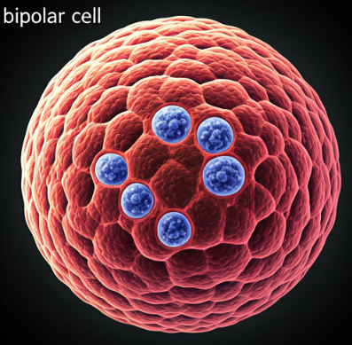
Bipolar cells are located in the inner nuclear layer of the eye. They work as intermediaries in the visual pathway, connecting the outer retina to the inner retina, which then sent the information to the brain, enabling visual perception.
Bipolar cells form a vital link between the outer layer and inner layer. Their primary function is to process and relay visual information received from photoreceptor cells to ganglion cells. This transmission is essential for image creation. Just like most neurons in the human eye, bipolar cells do not fire action potentials to transmit signals. Instead, they release glutamate, a neurotransmitter, in a graded manner. This means the amount of glutamate released depends on the intensity of the signal received from the photoreceptors(rod and cones).
Types of Bipolar Cells:-
There are two important types of bipolar cells:
- Rod bipolar cells(RBs): Highly sensitive to dim light. These cells receive electrical signals from rod photoreceptors, which are responsible for vision in low-light conditions. Rod bipolar cells are highly sensitive to light but these cells are enabled to detect color.
- Cone bipolar cells(CBs): These cells receive signals from cone photoreceptors, and pass light signals to other cells which are responsible for color vision in bright light. There are different subtypes of cone bipolar cells, each associated with specific types of cones (red, green, or blue), an eye contains 6 million cones.
Importance:-
Bipolar cells are essential for sight. These cells are the crucial link between the photoreceptor cells that detect light and the ganglion cells that send information to the brain. They also play an important role in processing visual information, ensuring we see things
clearly and accurately. Without these crucial middlemen, the information from the photoreceptor cells wouldn’t reach the brain. It would be like having a phone with no service. You could have the best camera in the world, but you would not be able to capture or send pictures.
Photorecepter Cells:-
Photoreceptor cells are special neurons in the retina, the primary function of these cells is to convert light into electrical signals, playing the first step in vision.
These cells are responsible for color and night vision. Imagine your eye as a high-tech camera. The lens focuses the light, and the film that captures the image is your retina, at the back of your eye. Now, on this film are special cells called photoreceptors. They are like the tiny pixels that record the light.
Types of Photoreceptor Cells:-
There are two main types of cells:
- Rods: Highly sensitive to low light and important for (night vision), detecting shades of gray. An eye contains 100 million rod cells.
- Cones: Responsible for color vision and sharp and bright light. An eye contains 6 million cones. The following list demonstrates the subtypes of cones.
- (S-cone short wavelength) They are specialized for detecting short wavelength light, responsible for color vision in the blue range.
- (M-cone medium wavelength) medium wavelength cones in the retina responsible for detecting green light.
- (L-cone large wavelength) plays a significant role in bright light, specialized for detecting long wavelengths of light, associated with red color discrimination. These wavelengths of the eye allow us to see multiple colors. An eye contains 6 million cones.
Importance:-
Photoreceptor cells are essential for vision. Without them, we would be blind. Damage to these cells, such as in macular degeneration or retinitis pigmentosa, can lead to significant vision loss or blindness.

